Some Asthma Symptoms Are Only Present During An Asthma Attack
An asthma attack is when a persons asthma symptoms become worse or more noticeable. During an attack, the muscles around the airways tighten more than usual, and the airways produce an overabundance of .
The typical signs of an asthma attack can include any of the following:
Wheezing This refers to a whistling or squeaky, almost musical sound during breathing.
Shortness of Breath This simply means feeling like you cant get enough air into your lungs.
Rapid Breathing In response to not getting enough air in each breath, your body may speed up your rate of breathing.
Coughing A during an asthma attack may contain .
Chest Tightness This can take the form of pain, pressure, or feeling like something is squeezing or sitting on your chest.
Not everyone with asthma experiences symptoms the same way, and asthma symptoms can differ between attacks. Asthma attacks require immediate treatment with a rescue or quick-relief or other medication recommended by your doctor.
How Is Copd Treated
While there is no cure for COPD, your doctor may recommend one or more of the following to help relieve symptoms:
- Lifestyle changes: Stop all smoking and increase physical activity.
- Therapies: Oxygen therapy involves the use of a device that brings additional oxygen to your lungs. Pulmonary rehabilitation is a program that uses counseling, diet advice and physical activities to help you better manage your COPD.
- Medications: Steroids, inhalers and antibiotics may be prescribed in an effort to treat symptoms of COPD.
- Surgery: In severe cases, major surgery, such as a lung or lung volume reduction surgery, may be needed when symptoms have not improved by way of medication or non-invasive therapies.
What Does S Chest X Ray Show
chest XrayshowslungChest XrayschestChest Xrays canlung
What is a chest x ray used to diagnose?
Xray Chest. It is used to evaluate the lungs, heart and chest wall and may be used to help diagnose shortness of breath, persistent cough, fever, chest pain or injury. It also may be used to help diagnose and monitor treatment for a variety of lung conditions such as pneumonia, emphysema and cancer.
can chest xray show heart attack?heart attackwillChest X-rayX-raychestheart
Contents
Don’t Miss: Can You Join The Army If You Have Asthma
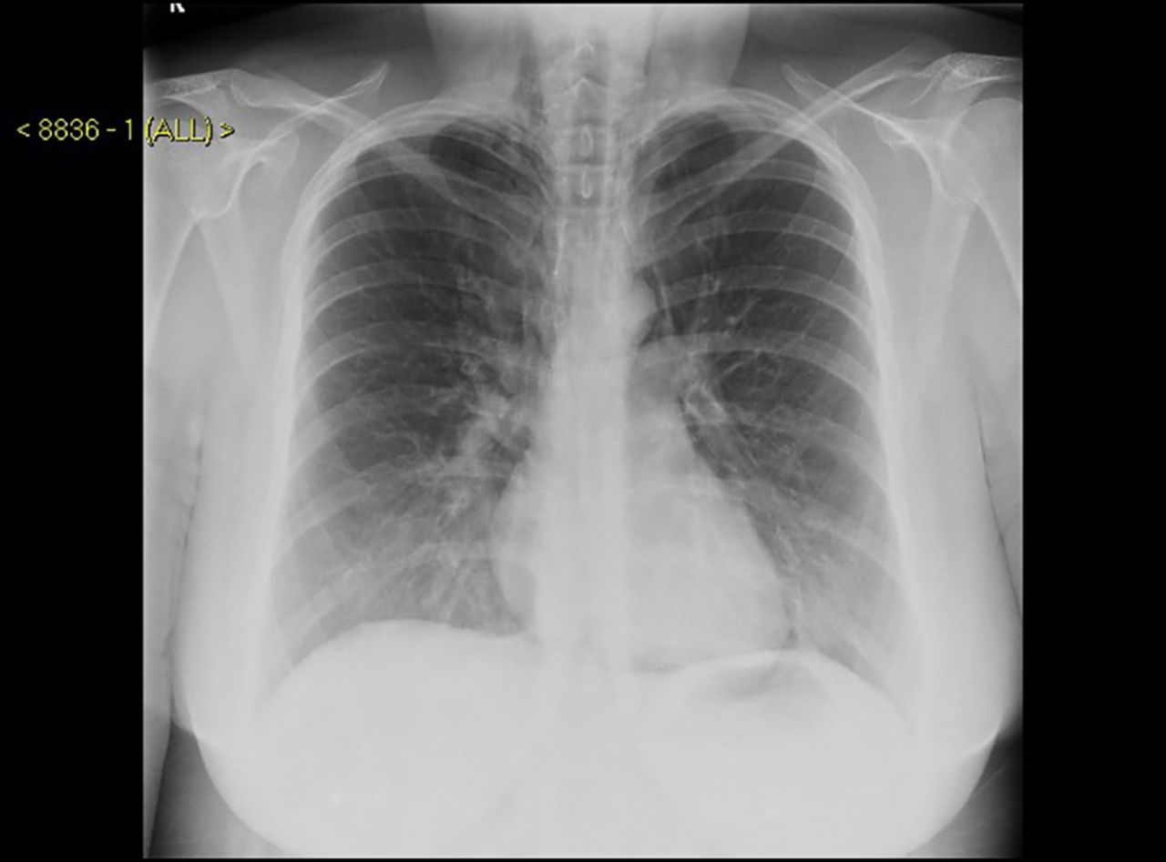
What Shows Up On A Chest X Ray
Chest Xrays can also reveal fluid in or around your lungs or air surrounding a lung. The image helps your doctor determine whether you have heart problems, a collapsed lung, pneumonia, broken ribs, emphysema, cancer or any of several other conditions.
Do X rays show tumors?
These include diagnosing tumors or bone injuries. Xrays are made by using external radiation to produce images of the body, its organs, and other internal structures for diagnostic purposes. A bone or a tumor, which is denser than soft tissue, allows few of the Xrays to pass through and appears white on the Xray.
What Is A Chest X
An X-ray is a type of screening test that takes a photographic or digital image of the structures inside the body. It is a painless and fairly quick screening that passes X-ray beams through the body to be absorbed to different degrees by different materials. X-rays hold a very small risk for radiation exposure .
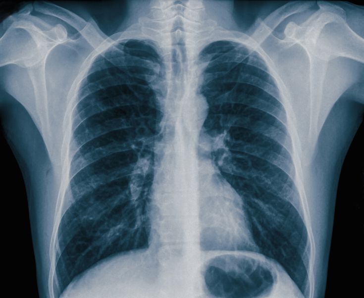
A chest X-ray points the X-ray beams towards the chest to take a picture of your lungs and chest area. A chest X-Ray shows:
- Lungs
- Several major blood vessels in the chest
- Ribs
- The air in your lungs
- Fat and muscle
Recommended Reading: Can Asthma Symptoms Last For Weeks
How Does Chest Infection Show On X Ray
5/5Chest Xrays caninfectioncanshowgiven here
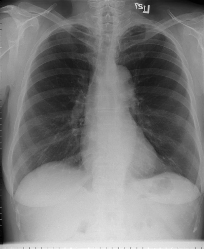
Chest x–ray: An x–ray exam will allow your doctor to see your lungs, heart and blood vessels to help determine if you have pneumonia. When interpreting the x–ray, the radiologist will look for white spots in the lungs that identify an infection.
Additionally, what does a shadow on a chest X ray mean? The white shadows on chest X–ray represent more dense or solid tissues, such as bone or heart, and the darker shadows on the chest X–ray represent air filled tissues, such as lungs.
Furthermore, how do doctors know if you have a chest infection?
Signs and symptoms of a chest infectioncoughing up yellow or green phlegm , or coughing up blood. breathlessness or rapid and shallow breathing. wheezing. a high temperature
What is a chest X ray used to diagnose?
A chest X–ray is a fast and painless imaging test that uses certain electromagnetic waves to create pictures of the structures in and around your chest. This test can help diagnose and monitor conditions such as pneumonia, heart failure, lung cancer, tuberculosis, sarcoidosis, and lung tissue scarring, called fibrosis.
How Do You Feel When You Have Asthma
All of these factors bronchospasm, inflammation, and mucus production cause symptoms such as difficulty breathing, wheezing, coughing, shortness of breath, and difficulty performing normal daily activities. Other symptoms of an asthma attack include: Severe wheezing when breathing both in and out.
Read Also: Does Ibuprofen Make Asthma Worse
What Happens During A V/q Scan
For the ventilation part of the scan you will be asked to breathe in a slightly radioactive gas called krypton for about 10 minutes. This is odourless and tasteless.
For lung perfusion you will be given an injection of slightly radioactive material. Sometimes only the perfusion part of the scan is needed.
The whole process usually takes up to an hour.
Do Not Routinely Order Chest X
There is extensive evidence that the majority of X-rays ordered for children admitted for asthma and wheezing disorders do not provide clinically relevant information and therefore do not contribute to their diagnosis and management.
Clear clinical criteria outlining the indications for X-rays in asthma should be defined to avoid unwarranted chest X-rays in children with acute wheeze.
Supporting evidence
- Hederos C-A, Janson S, Andersson H, et al. Chest x-ray investigation in newly discovered asthma. Pediatric Allergy and Immunology 2004 15: 163165.
- Muthukrishnan L, Raman R. Analysis of clinical & radiological findings in children with acute wheeze. Pulmonary and Respiratory Research 2013 1:1
- Narayanan S, Magruder T, Walley SC, et al. Relevance of chest radiography in pediatric inpatients with asthma. Journal of Asthma 2014 51:751-5.
The Paediatrics & Child Health Division formed a group of interested Fellows to comprise a General Paediatrics EVOLVE Working Group. A review of low-value practices relevant to general paediatrics was conducted drawing on lists published by Choosing Wisely US and Canada, contributions to Choosing Wisely Australia by other medical colleges and published EVOLVE lists developed by other specialties in order to identify low-value practices of relevance while avoiding duplicating the mention of practices already identified in other EVOLVE lists. Based on this review, the Working Group shortlisted 15 items for further consideration.
Also Check: What Two Body Systems Are Affected By Asthma
What Are Common Ways To Diagnose Asthma
Personal and medical history. Your doctor will ask you questions to understand your symptoms and their causes. Bring notes to help jog your memory. Be ready to answer questions about your family history, the medicines you take and your lifestyle. This includes any current physical problems. Shortness of breath, wheezing, coughing and tightness in your chest may show asthma. This also includes all previous medical conditions. A history of allergies or eczema increases your chance of asthma. A family history of asthma, allergies or eczema increases your chance of having asthma, too. Tell your doctor about any home or work exposure to environmental factors that can worsen asthma. For example, these might include pet dander, pollen, dust mites and tobacco smoke. The doctor may also ask if you get chest symptoms when you get a head cold.
Physical exam. If your doctor thinks you have asthma, they will do a physical exam. They will look at your ears, eyes, nose, throat, skin, chest and lungs. This exam may include a lung function test to detect how well you exhale air from your lungs. You may also need an X-ray of your lungs or sinuses. A physical exam then allows your doctor to review your health.
When To See A Specialist About Your Asthma
Asthma is not always easy to diagnose, Fineman says, but you should see your doctor if youre having repeated episodes of wheezing and coughing or shortness of breath. If youre diagnosed with the condition, work with your doctor to develop an asthma management and action plan.
Although your primary care doctor may be able to diagnose and treat your asthma, if your symptoms dont respond to a first-line therapy of inhaled and short-acting bronchodilators, Asciuto recommends that you see a lung specialist or allergy and asthma specialist.
You May Like: Albuterol And Ibuprofen
How Can I Prepare For A Chest X
Youll usually receive an appointment letter. Read this carefully in case there is anything special you need to do to prepare. The main thing you need to do is to take off anything metal, such as jewellery, zips or belt buckles.
If you could be pregnant, its important to tell the radiographer, so that they can reduce any exposure of your unborn baby to X-rays.
At The Specialist Asthma Clinic
If youre taking your asthma medicines exactly as prescribed and using the right inhaler technique, but youre still getting symptoms, your GP or asthma nurse may refer you to a specialist asthma clinic. It may take some time before you get an appointment, so youll still have to manage your condition through your GP until then.
At the clinic they will try to work out whats going on by answering these questions:
- Is the type of asthma you have difficult to control asthma?
- Is the type of asthma you have severe asthma?
- Is there another reason why youre getting your asthma symptoms?
You might have had tests to diagnose and monitor your asthma already, but to confirm or rule out a diagnosis of severe asthma you may need some extra tests.
You probably wont need all these tests, though. Your asthma specialist will explain which ones you need and why.
Everyone with asthma is different so the tests youll need will depend on your individual asthma symptoms, medical history, family history and any other conditions you have, says Asthma UKs in-house GP Dr Andy Whittamore.
And because your asthma symptoms can vary over time, you may need to have these tests more than once to help your asthma specialist make the right diagnosis.
Don’t Miss: What’s An Asthma Attack Feel Like
Breathing Problems During Exercise
If you have chest tightness, cough, wheeze or shortness of breath during exercise, your doctor may perform extra tests to see if you have a type of asthma called, exercise-induced asthma or exercise-induced bronchospasm. For some people, they will only have asthma symptoms during exercise. There are many benefits to exercise, so work with your doctor to find the best management steps and treatment options for you.
What Happens During A Pet Scan
PET scanners have a flat bed with a large, circular scanner at one end. Before a scan, youll be injected with a slightly radioactive substance, which can be detected by the scanner. You will be asked to lie down on the table which will slide into the PET scanner. Its important that you stay as still as you can while you are in the scanner. PET scans are painless and take around 3060 minutes.
Also Check: Omeprazole And Asthma
Racp Paediatrics & Child Health Division
Recommendations from the RACP’s Paediatrics & Child Health Division on antibiotic use, bronchiolitis, asthma diagnosis, GORD treatment and abdominal x-rays.The Paediatrics & Child Health Division represents 4,500 Fellows and Trainees of The Royal Australasian College of Physicians . We aim to improve the health and wellbeing of neonates, infants and children as well as adolescents and young adults through education and training, research, and policy and advocacy.
What Is A Chest Ct Scan
Unlike a traditional X-ray, a chest CT creates 2-dimensional, cross-sectional images. The series of computerized views taken from different angles create detailed pictures of your chest area. The computer collects the images and arranges them in order for your doctor to view.4
For a CT scan, you will be instructed to lie flat on the CT scan table. You will move quickly through a scanner, which is a doughnut-shaped device. You may be told to lie still and hold your breath for a few seconds. A CT scanner is open and much less noisy than an MRI, and a CT scan takes less time. If your doctor wants the CT scan done with contrast, you will get an intravenous injection. This may give you a warm feeling throughout your body, but it is just temporary. At the end of the scan, you can go about your activities as usual.4
You May Like: Does Ibuprofen Make Asthma Worse
Does Laying Down Help Asthma
Good sleep equals healthy and clear air passages. Yet, even gravity has an effect on night-time breathing. When we lie down flat, our chest area naturally collapses into a more relaxed state but this can put pressure on the lungs. If it’s comfortable for you, try propping yourself up on a higher pillow as you sleep.
Why Would I Need A Chest Ct For Asthma
A chest CT scan is currently the gold standard to make a diagnosis of asthma, as well as to look for any complications. If you have chronic asthma symptoms or if your symptoms keep recurring, this scan can help doctors pinpoint a diagnosis.1
For example, a CT scan can be helpful in diagnosing allergic bronchopulmonary aspergillosis , a condition associated with asthma. ABPA is most common in people with longstanding asthma. It has symptoms that are very similar to asthma, like wheezing, shortness of breath, weight loss, and fatigue.5-7
A CT scan can also be helpful in detecting health conditions that can seem like asthma. Another possible condition that your doctor may want to rule out with a CT scan is . Bronchiectasis causes your bronchial tubes to become thickened from inflammation. It can cause periodic flare-ups of breathing difficulties.5
CT scans are used to check for complications of asthma and to rule out other health conditions. In the future, these and other imaging scans may also be useful in providing personalized asthma care.1
Recommended Reading: Warm Or Cool Mist Humidifier For Asthma
What A Chest X
In early-stage disease, a chest X-ray may, in fact, appear quite normal. This doesn’t mean that there is no damage it is simply that the test has limitations as to how much it can visually tell us. It can neither describe your individual lung capacity nor the force by which you can inhale or exhale air.
What it can do is give us a visual reference point by which to compare any changes that may develop over time. As such, doctors will typically recommend a chest X-ray every one or two years depending on how far along your COPD is.
In later-stage disease, visual changes will become more apparent. One of the most obvious features will be the so-called hyperinflation of the lungs. When this happens, the doctor will be able to see several things on the X-ray:
- A flattening of the diaphragm as the lungs press down on the muscle
- Increased chest size as measured from front to back
- An elongated and narrow heart
- Pockets of air called bullae around a half inch in size or larger
In the event your doctor needs a more extensive view of the lung structure and damage, a computed tomography scan may be ordered. Where a chest X-ray will only deliver a one-dimensional image of the lungs, a CT scan will take a series of images to create a more three-dimensional representation. In doing so, the CT scan can pick up finer detail and provide doctors a more complete portrait of the person’s COPD.
Why A Chest X
- Posted on: Jun 15 2018
- Leave a response
The x-ray is one of the most common imaging methods performed in hospitals and imaging centers like ours. It is widely understood that this type of radiology enables doctors to observe hard tissue like bones. Additionally, there is value in x-ray imaging for the chest cavity. Here, we discuss a few of the reasons why a chest x-ray may be ordered.
Don’t Miss: How To Get Rid Of Asthma Without Inhaler
Other Tests You May Need If You Have Asthma
Even if your lung function tests are normal, your doctor may order other tests to see what could be causing your asthma symptoms.
- Gas and diffusion tests can measure how well your absorbs oxygen and other gases from the air you breathe. You breathe in a small amount of a gas, hold your breath, then blow out. The gas you exhale is analyzed to see how much your blood has absorbed.
- X-rays may tell if there are any other problems with your lungs, or if asthma is causing your symptoms. High-energy radiation creates a picture of your lungs. You may be asked to briefly hold your breath while you stand in front of the X-ray machine.