What Are The Risks Of A Chest X
Chest X-rays expose the patient briefly to a minimum amount of radiation. Any radiation exposure has some risk to the tissues of the body. The radiation exposure in a chest X-ray is minimized by the type of X-ray high-speed film, which does not require as much radiation exposure as in the past. The radiology technician is guided by technique standards which have been established by national and international guidelines. These guidelines are designed and reviewed by both the Department of Health and Human Services and national and international radiology protection councils.
Women who are , especially in early , should notify their physicians, as the fetus is at risk for harm with any radiology technique. X-rays are typically avoided in pregnant patients unless absolutely necessary, in which case the patient’s abdomen is covered with a special lead gown to block the radiation from the fetus.
What To Expect When Having A Chest X
X-rays are usually taken by a trained and certified radiology technician. Patients who are undergoing an X-ray of the chest will put on a special gown and remove all metallic items, including jewelry so that they don’t block the X-ray beam from penetrating the body.
The X-ray technician may ask the patient to inhale deeply and hold her breath during the procedure to inflate the lungs and make the various chest tissues more visible. X-rays may be taken from the front, back and side views, and from different camera angles while sitting, standing or lying down.
Once the X-ray has been taken, the exposed film is placed into a developing machine and the image is examined and interpreted by a radiologist . After the radiologist reviews the X-ray, he or she will send a report to the doctor who ordered the test. This doctor will then discuss the results and recommended treatment options with the patient.
The risks of chest X-rays are minimal, especially because today’s high-speed film does not require as much radiation exposure as the type of film used years ago. However, any exposure to radiation has some risk, which is why the technician asks the patient to wear a lead apron over the reproductive parts of the body or the extremities to shield from exposure. Pregnant women should ask their physicians before having an X-ray taken, as this could harm the fetus.
What Is A Chest X
An X-ray is a type of screening test that takes a photographic or digital image of the structures inside the body. It is a painless and fairly quick screening that passes X-ray beams through the body to be absorbed to different degrees by different materials. X-rays hold a very small risk for radiation exposure .
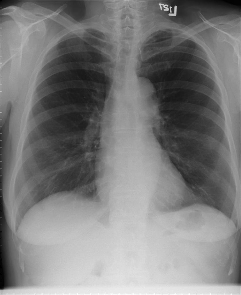
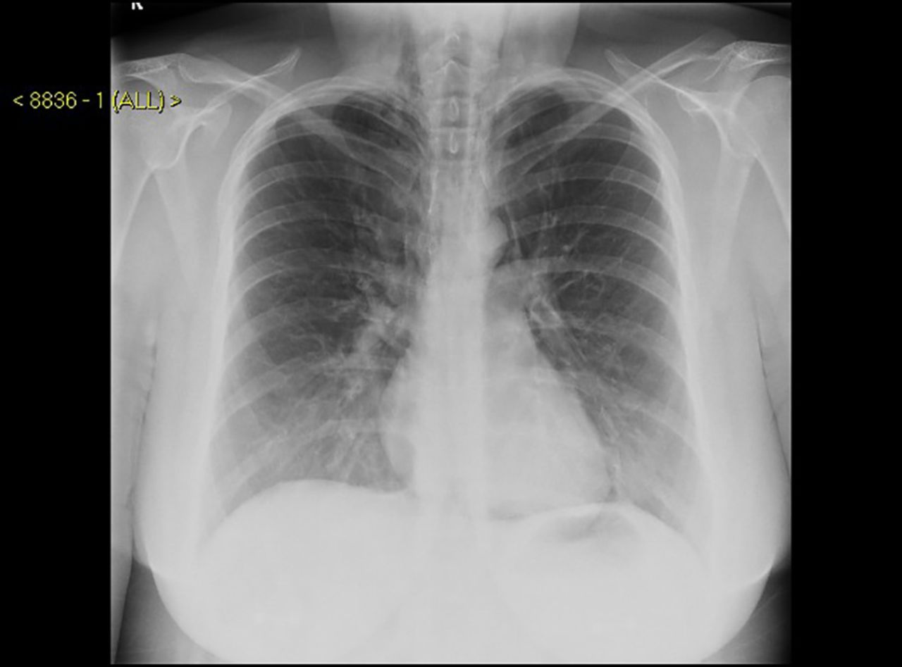
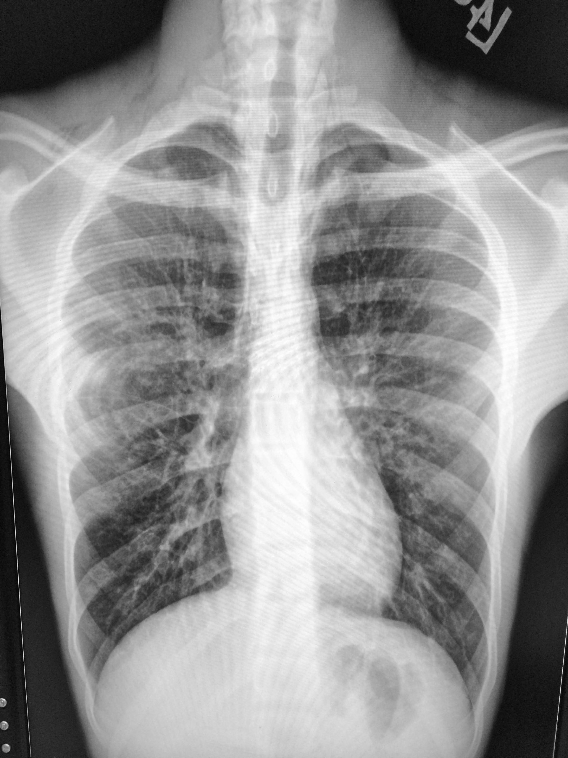
A chest X-ray points the X-ray beams towards the chest to take a picture of your lungs and chest area. A chest X-Ray shows:
- Lungs
- Several major blood vessels in the chest
- Ribs
- The air in your lungs
- Fat and muscle
Bronchiectasis And Bronchial Dilatation
Studies of HRCT images in asthma consistently reveal the presence of bronchiectasis in patients with asthma but not allergic bronchopulmonary aspergillosis . In ABPA, bronchiectasis often is considered part of the definition of the disease. Dilated airways may take the form of cylindrical, varicose, or cystic bronchiectasis. Park et al observed bronchial dilatation in 31% of patients with asthma versus 7% of control subjects. The authors measured bronchoarterial ratios but did not find a statistically significant difference between the groups.
Lynch et al showed that dilated bronchi, defined as bronchi that are larger than accompanying arteries in which the tapering pattern is not lost, were observed in 59% of the control subjects, as compared to 77% of the patients with asthma. Other researchers found no or few such features in control subjects. A decreased arterial diameter with hypoventilation and hypoxic vasoconstriction, a sectioning artifact near the branching arteries and bronchi, a bronchodilator effect on medium-sized airways, and subclinical ABPA are potential explanations for the unexpectedly high percentage of findings in control subjects.
In a study by Lo et al of 30 children with difficult-to-treat asthma, abnormal CT findings were highly prevalent in a cohort of children with severe asthma, with bronchiectasis identified in approximately 27%. Bronchial wall thickening was observed in 80%, and air trapping in 60%.
Breathing Problems During Exercise

If you have chest tightness, cough, wheeze or shortness of breath during exercise, your doctor may perform extra tests to see if you have a type of asthma called, exercise-induced asthma or exercise-induced bronchospasm. For some people, they will only have asthma symptoms during exercise. There are many benefits to exercise, so work with your doctor to find the best management steps and treatment options for you.
Some Asthma Symptoms Are Only Present During An Asthma Attack
An asthma attack is when a persons asthma symptoms become worse or more noticeable. During an attack, the muscles around the airways tighten more than usual, and the airways produce an overabundance of .
The typical signs of an asthma attack can include any of the following:
Wheezing This refers to a whistling or squeaky, almost musical sound during breathing.
Shortness of Breath This simply means feeling like you can’t get enough air into your lungs.
Rapid Breathing In response to not getting enough air in each breath, your body may speed up your rate of breathing.
Coughing A during an asthma attack may contain .
Chest Tightness This can take the form of pain, pressure, or feeling like something is squeezing or sitting on your chest.
Not everyone with asthma experiences symptoms the same way, and asthma symptoms can differ between attacks. Asthma attacks require immediate treatment with a rescue or quick-relief or other medication recommended by your doctor.
How Is Copd Treated
While there is no cure for COPD, your doctor may recommend one or more of the following to help relieve symptoms:
- Lifestyle changes: Stop all smoking and increase physical activity.
- Therapies: Oxygen therapy involves the use of a device that brings additional oxygen to your lungs. Pulmonary rehabilitation is a program that uses counseling, diet advice and physical activities to help you better manage your COPD.
- Medications: Steroids, inhalers and antibiotics may be prescribed in an effort to treat symptoms of COPD.
- Surgery: In severe cases, major surgery, such as a lung or lung volume reduction surgery, may be needed when symptoms have not improved by way of medication or non-invasive therapies.
Other Tests For Conditions That May Mimic Asthma
There are some medical conditions that often make asthma harder to treat and control, in addition to being asthma mimics. These include allergies and GERD. If you are diagnosed with asthma, your doctor might also test you for these conditions, or treat them for several weeks to see if your asthma symptoms also improve.
For more information on allergies, GERD, and other triggers, see Causes of Asthma.
What Is A Chest Radiograph
Also known as a chest X-ray, a chest radiograph uses an X-ray beam. It is considered the best general technique for looking at the lungs, the chest cavity, and the area around the lungs.1,2
To undergo a chest X-ray, you will typically stand in front of a wall-mounted device that holds the X-ray film or a plate that digitally records the image. For the first view, you will probably be asked to stand with your hands on your hips, with your chest pressed against the image plate. For a second view, you may be asked to elevate your arms.3
During the procedure, the X-ray machine emits a small burst of radiation that goes through the body, recording an image. The entire procedure takes only about 15 minutes. When you are finished, you may be asked to wait until the radiologist lets you know that all the needed images have been obtained.3
The majority of people with asthma will have a chest radiograph. However, if you have difficult-to-treat or severe asthma, then a CT scan may be ordered.
Use Of Imaging In The Clinical Evaluation Of Asthma
Imaging of the lungs in patients with asthma has evolved dramatically over the last decade; however, the clinical diagnosis of asthma is still based on a compatible history, exam findings and evidence of variable airflow obstruction. Chest imaging is most helpful in evaluating complications from asthma and ruling out alternative diagnoses. The chest radiograph findings are non-specific and often may be normal. The most common abnormal finding is bronchial wall thickening, present in 48-71% of radiographs , followed by hyperinflation found in 24% of cases in one series . Marked hyperinflation is most often seen in the setting of emphysema. Previous studies evaluating the need for chest radiographs in acute asthma exacerbations revealed that patients presenting to the emergency department with uncomplicated asthma have abnormal chest x-rays only 1-2.2% of the time . However, this number increases to nearly 34% in patients who are unresponsive to initial bronchodilator therapy and require admission to the hospital . Abnormalities that may change management include pneumothorax, pulmonary vascular congestion, focal parenchymal opacities, and enlarged cardiac silhouette.
CT chest lung densityCT chest 3D bronchial tree
Figure demonstrates labeling of the bronchial tree out to the segmental bronchi of a subject with severe asthma enabling each segmental bronchial wall thickness to be measured quantitatively. Image processing derived from Apollo software .
How Should A Normal Chest X Ray Look
4.8/5Chest Xraychest radiographchestCXRChest Xraychestrelated to it here
Chest X–rays can detect cancer, infection or air collecting in the space around a lung . They can also show chronic lung conditions, such as emphysema or cystic fibrosis, as well as complications related to these conditions. Heart-related lung problems.
Secondly, can a chest xray show COPD? While a chest x-ray may not show COPD until it is severe, the images may show enlarged lungs, air pockets or a flattened diaphragm. CT is sometimes used to measure the extent of emphysema within the lungs. It can also help determine if the symptoms are the result of another disease of the chest.
In this regard, what does a chest X ray look like with pneumonia?
When interpreting the x–ray, the radiologist will look for white spots in the lungs that identify an infection. CT of the lungs: A CT scan of the chest may be done to see finer details within the lungs and detect pneumonia that may be more difficult to see on a plain x–ray.
Can chest xray show cancer breast?
While chest X-rays have a low success rate in detecting whether breast cancer has spread to your lungs, your doctor may still recommend one for several reasons.
How Vets Treat Asthma In Cats
Cats can have asthmatic symptoms for a single occurrence, a short time, a few years or an entire lifetime. The important goal is to be prepared for future attacks.
While asthma cant be cured, it can be successfully managed. This can be achieved by working with your vet or respiratory specialist and following the instructions given to you.
A cat having an asthma attack needs intravenous steroids to desensitize the airways and reverse inflammation.
Inhalers are also a great way to manage the condition, although getting the cat to breathe in the medication can be tricky, even with specially designed dosing chambers.
I remember talking to an eminent internal medicine specialist with an asthmatic cat. As is typical with many animals belonging to veterinarians, this cat refused to take medication , and so her mom was forced to give a less sophisticated treatment of once-monthly depot injections of steroid.
Long-term steroids are extremely effective at suppressing inflammation.
Depression And Anxiety Scores

These questionnaires are used to find out how having severe asthma is affecting you emotionally. In an Asthma UK survey, 68% of people with severe asthma said theyd had anxiety and 52% of people with severe asthma said they felt depressed.
Because asthma symptoms that affect your quality of life can be worrying and get you down, its important for your healthcare team to understand how youre feeling, says Dr Andy.
If youre seeing an asthma specialist to diagnose or rule out severe asthma, they will usually consider screening you for anxiety and depression often using these questionnaires. For some people with asthma, treating the depression and anxiety can have a big impact on your symptoms and how they affect you.
Evidence shows that people with low mood or anxiety can find it harder to control their asthma symptoms, says Dr Andy. Its important that your asthma specialist and healthcare team have a good understanding of how youre feeling so they can give you the support you need.
Next review due March 2022
Why Would I Need A Chest Ct For Asthma
A chest CT scan is currently the gold standard to make a diagnosis of asthma, as well as to look for any complications. If you have chronic asthma symptoms or if your symptoms keep recurring, this scan can help doctors pinpoint a diagnosis.1
For example, a CT scan can be helpful in diagnosing allergic bronchopulmonary aspergillosis , a condition associated with asthma. ABPA is most common in people with longstanding asthma. It has symptoms that are very similar to asthma, like wheezing, shortness of breath, weight loss, and fatigue.5-7
A CT scan can also be helpful in detecting health conditions that can seem like asthma. Another possible condition that your doctor may want to rule out with a CT scan is . Bronchiectasis causes your bronchial tubes to become thickened from inflammation. It can cause periodic flare-ups of breathing difficulties.5
CT scans are used to check for complications of asthma and to rule out other health conditions. In the future, these and other imaging scans may also be useful in providing personalized asthma care.1
At The Specialist Asthma Clinic
If youre taking your asthma medicines exactly as prescribed and using the right inhaler technique, but youre still getting symptoms, your GP or asthma nurse may refer you to a specialist asthma clinic. It may take some time before you get an appointment, so youll still have to manage your condition through your GP until then.
At the clinic they will try to work out whats going on by answering these questions:
- Is the type of asthma you have difficult to control asthma?
- Is the type of asthma you have severe asthma?
- Is there another reason why youre getting your asthma symptoms?
You might have had tests to diagnose and monitor your asthma already, but to confirm or rule out a diagnosis of severe asthma you may need some extra tests.
You probably wont need all these tests, though. Your asthma specialist will explain which ones you need and why.
Everyone with asthma is different so the tests youll need will depend on your individual asthma symptoms, medical history, family history and any other conditions you have, says Asthma UKs in-house GP Dr Andy Whittamore.
And because your asthma symptoms can vary over time, you may need to have these tests more than once to help your asthma specialist make the right diagnosis.
When To See A Specialist About Your Asthma
Asthma is not always easy to diagnose, Fineman says, but you should see your doctor if youre having repeated episodes of wheezing and coughing or shortness of breath. If you’re diagnosed with the condition, work with your doctor to develop an asthma management and action plan.
Although your primary care doctor may be able to diagnose and treat your asthma, if your symptoms dont respond to a first-line therapy of inhaled and short-acting bronchodilators, Asciuto recommends that you see a lung specialist or allergy and asthma specialist.
Symptoms Of Asthma In Cats
Asthma affects the lower airways and causes narrowing of the bronchi, increased mucus production and decreased mucus clearance. The end result is a cat who struggles to breathe.
Signs of asthma in a cat can be minor to severe. A dry, hacking cough is most often mistaken for a hairball, and this coughing may be occasional or daily.
Additional symptoms may include:
- Open-mouthed panting or breathing
- Hunched on the ground/floor with neck extended
- Lips and gums appear blue
A coughing cat is a rare thing and is commonly linked with feline asthma.
However, the absence of a cough does not rule asthma out. During an asthma attack, many cats have a wheeze that you can hear from across the room but again, not all cats do. However, a cat in the grip of an asthma attack does have problems breathing.
Signs of difficult breathing include the cat sitting in the same spot for ages to rest and concentrate on breathing. They extend their head and neck into a straight line and open their mouth , and their chest moves in and out in an exaggerated manner.
Sometimes, the cats airways are so narrow that they have to use their stomach muscles to push air out of their lungs to get ready for the next breath this is known as abdominal effort.
A desperately ill cat has gums tinged lilac or blue instead of a healthy pink. If you suspect your cat is having difficulty breathing , never stress them. Leave them resting while you contact the vet and follow the vets instructions.
Symptom And Quality Of Life Scores
These tests involve filling out questionnaires to find out what symptoms youre experiencing and the different ways asthma may be affecting your life.
In an Asthma UK survey, 91% of people said their severe asthma had an impact on everyday things, such as their ability to exercise, their family and social life, their work or school life and their holidays. In the same survey 82% of people said theyd experienced sleep loss and 66% had gained weight.
If your asthma symptoms mean you cant do things you used to be able to do, such as exercise or play with your children, these scores will help your asthma specialist find out how asthma is affecting your life, says Dr Andy. Theyre also a useful way to monitor how well your asthma treatments are working.
Asthma specialists use these tests to:
- give them a clearer picture of your asthma symptoms
- show them how asthma may be limiting your activities
- reveal how youre coping with the challenges of having frequent asthma symptoms
- work out your response to longer-term treatment trials.
Imaging As A Biomarker
The effect of inhaled corticosteroid use on air trapping in mild to moderate asthma patients with uncontrolled symptoms has been assessed using CT. After completing 3 months of therapy with an inhaled corticosteroid, patients exhibited a decrease in air trapping as measured by CT . Thus, air trapping can serve as a potential outcome related to disease control. Recently, biologic therapy with anti-IL5 monoclonal antibody has shown promise to reverse airway remodeling process. Haldar et al demonstrated in 26 patients with severe refractory asthma with sputum eosinophilia that treatment with mepolizumab significantly decreased average wall area over one year compared with placebo . Current analysis tools allow for measurement of the ventilation defect percentage from 129Xe and 3He MRI, which is the volume of lung not involved in ventilation. Texture features can also be generated from MRI ventilation images and can be used to quantify differences in lung ventilation post bronchodilator therapy . Further studies are needed to evaluate the optimal imaging biomarker to assess response to biologic therapy in patients with asthma.
Who Can Interpret Chest X
Many doctors are trained to interpret chest X-rays. In addition to radiologists, who have special training in reading all radiology films, primary care physicians, internists, pediatricians, emergency room doctors, anesthesiologists, heart doctors , lung doctors and lung surgeons are the doctors who frequently interpret chest X-rays as a part of their routine practice.
What Is Nipple Shadow Chest Xray

Nipple shadowsnippleschest
Nipple shadows are often seen on chest x-rays and can be easily confused for a pulmonary nodule or nodules. If there is any doubt the easiest method of determining whether opacities represent nipple shadows is a repeat chest x-ray with nipple markers.
Likewise, what does it mean if you have a shadow on your lung? Pulmonary edema is a condition involving the accumulation of fluid in the lungs, often due to heart disease. Aortic aneurysm can cause a shadow on chest X-rays.
Just so, what is a chest xray with nipple markers?
and Dr Ian Bickle ? et al. Nipple markers can be a useful technique in the evaluation of small radiodensities overlying the expected position of the nipple on a chest radiograph. Often, especially in women, this is a nipple shadow – a dense nipple projected over the lung.
Are lung nodules always cancer?
Yes, lung nodules can be cancerous, though most lung nodules are noncancerous . Lung nodules small masses of tissue in the lung are quite common. They appear as round, white shadows on a chest X-ray or computerized tomography scan.
Other Tests You May Need If You Have Asthma
Even if your lung function tests are normal, your doctor may order other tests to see what could be causing your asthma symptoms.
- Gas and diffusion tests can measure how well your absorbs oxygen and other gases from the air you breathe. You breathe in a small amount of a gas, hold your breath, then blow out. The gas you exhale is analyzed to see how much your blood has absorbed.
- X-rays may tell if there are any other problems with your lungs, or if asthma is causing your symptoms. High-energy radiation creates a picture of your lungs. You may be asked to briefly hold your breath while you stand in front of the X-ray machine.
The Respiratory System: How It Should Work
Respiration is the process by which our bodies inhale oxygen and express carbon dioxide. This process becomes more difficult during an asthmatic attack.
When you inhale, air passes through your windpipe . Meanwhile your diaphragm contracts and moves downward creating air space in your chest cavity. The air enters your lungs, passing through the bronchial tubes and finally into tiny air sacs .
Oxygen from the air passes from the alveoli and into the bloodstream through tiny blood vessels called capillaries. Capillaries deliver this oxygen-rich blood to pulmonary veins, which pass it to the left side of your heart. The heart then pumps the oxygen-rich blood to the rest of your body.
When you exhale, air that is rich in carbon dioxide passes out of your lungs, through your windpipe, and out your body through your nose and/or mouth.
Disrupted Sleep Difficulty Exercising And Some Other Signs Can Indicate You Might Have Asthma
Along with its short-term symptoms, asthma can cause other problems or disruptions.
Because symptoms often become worse at night, asthma can disrupt sleep or cause . Poor sleep, along with daytime asthma symptoms, can make it hard to complete work or school tasks, as well as day-to-day chores.
Asthma can make exercise challenging or impossible, which may put you at risk for a host of other medical problems.
RELATED: Complications That Can Occur With Asthma
Over time, if asthma is not properly treated or controlled with medication, it can cause airway remodeling, when the airways become scarred or permanently deformed. This can make breathing and treatment even more difficult.
Asthma is also associated with a greater risk for anxiety, depression, and other mental health disorders. If you see patterns of any of these problems or complications, its important to talk to your doctor to get to the root cause of the problem.
And remember, asthma symptoms do not look the same in everyone. Each person with asthma is unique and so are their symptoms, Dinakar says.
In some people with asthma, symptoms are very mild and seldom show up. In others, symptoms may be severe but situational, for example, after running hard or while going to bed. In others, symptoms are always around and may make everyday life difficult.
With additional reporting by Quinn Phillips and Markham Heid.
What Is A Chest Ct Scan
Unlike a traditional X-ray, a chest CT creates 2-dimensional, cross-sectional images. The series of computerized views taken from different angles create detailed pictures of your chest area. The computer collects the images and arranges them in order for your doctor to view.4
For a CT scan, you will be instructed to lie flat on the CT scan table. You will move quickly through a scanner, which is a doughnut-shaped device. You may be told to lie still and hold your breath for a few seconds. A CT scanner is open and much less noisy than an MRI, and a CT scan takes less time. If your doctor wants the CT scan done with contrast, you will get an intravenous injection. This may give you a warm feeling throughout your body, but it is just temporary. At the end of the scan, you can go about your activities as usual.4
