What Happens During A Pet Scan
PET scanners have a flat bed with a large, circular scanner at one end. Before a scan, youll be injected with a slightly radioactive substance, which can be detected by the scanner. You will be asked to lie down on the table which will slide into the PET scanner. Its important that you stay as still as you can while you are in the scanner. PET scans are painless and take around 3060 minutes.
Does An Inhaler Break Up Mucus
Techniques to remove mucus are often done after using an inhaled bronchodilator medication. The medication helps loosen the mucus and open the airways to make the techniques more effective. Common techniques used to help remove mucus include these, which can be ordered and demonstrated by your doctor.
What Are Some Common Chest X
Chest X-ray is generally used in combination with other clinical data such as, physical examination and the patient’s history and symptoms. It can also be used in combination of other radiology test to support, confirm, or exclude many conditions or diagnoses.
A chest X-ray can be used to define abnormalities of the lungs such as excessive fluid , fluid around the lung , pneumonia, bronchitis, asthma, cysts, and cancers. Heart abnormalities, including fluid around the heart , an enlarged heart , heart failure, or abnormal anatomy of the heart can be revealed on the films. Certain bony structures of the chest and broken bones or abnormalities of the bones of the spine in the chest can often be detected.
Also Check: Chihuahua Dogs And Asthma
At The Specialist Asthma Clinic
If youre taking your asthma medicines exactly as prescribed and using the right inhaler technique, but youre still getting symptoms, your GP or asthma nurse may refer you to a specialist asthma clinic. It may take some time before you get an appointment, so youll still have to manage your condition through your GP until then.
At the clinic they will try to work out whats going on by answering these questions:
- Is the type of asthma you have difficult to control asthma?
- Is the type of asthma you have severe asthma?
- Is there another reason why youre getting your asthma symptoms?
You might have had tests to diagnose and monitor your asthma already, but to confirm or rule out a diagnosis of severe asthma you may need some extra tests.
You probably wont need all these tests, though. Your asthma specialist will explain which ones you need and why.
Everyone with asthma is different so the tests youll need will depend on your individual asthma symptoms, medical history, family history and any other conditions you have, says Asthma UKs in-house GP Dr Andy Whittamore.
And because your asthma symptoms can vary over time, you may need to have these tests more than once to help your asthma specialist make the right diagnosis.
How Can I Prepare For A Pet Scan
Youll receive a letter from the hospital telling you how to prepare for your PET scan. Read it carefully. You’ll usually be advised not to eat anything for 6 hours beforehand.
Drinking fluid is allowed, but you should ideally just drink water. You should also avoid strenuous exercise for 24 hours before your appointment.
Its important to be on time for your PET scan. Thats because the radioactive substance used in the scan only has a short shelf life. Your scan may not work properly if its not done on time, and the test may be cancelled. If this happens, youll have to wait for another appointment.
Youll be told to avoid babies and children for a few hours after a PET scan, because you will be slightly radioactive. If you have children, its important to organise childcare to cover this time.
You May Like: Small Airways Inflammation
Tests For Other Conditions
The doctor may also do tests for other conditions that can make asthma worse, like:
- Gastroesophageal reflux disease
The Canadian Lung Association: “Signs and Symptoms of Asthma: Diagnosis.”
National Jewish Medical and Research Center: “How Is Asthma Diagnosed?”
American College of Allergy, Asthma & Immunology: “About Asthma: Diagnosing Asthma.”
Mayo Clinic: âAsthma: Steps in testing and diagnosis.â
Medscape: âAsthma Guidelines.â
American Academy of Asthma, Allergy, and Immunology: “What to expect at the doctor’s office.”
American Lung Association: “Spirometry and Other Lung Function Tests Fact Sheet.”
American Medical Association: Essential Guide to Asthma.
Asthma and Allergy Foundation of America: “Peak Flow Meters.”
Grayson, M. ACP Medicine, 2005.
National Asthma Education and Prevention Program: “Expert Panel Report 3: Guidelines for the Diagnosis and Management of Asthma — 2002.”
National Heart, Lung and Blood Institute: “Asthma: How is Asthma Diagnosed?”
MedlinePlus: “Pulmonary Function Tests.”
National Lung Health Education Program: “Spirometry.”
What Is Nipple Shadow Chest Xray
Nipple shadowsnippleschest
Nipple shadows are often seen on chest x-rays and can be easily confused for a pulmonary nodule or nodules. If there is any doubt the easiest method of determining whether opacities represent nipple shadows is a repeat chest x-ray with nipple markers.
Likewise, what does it mean if you have a shadow on your lung? Pulmonary edema is a condition involving the accumulation of fluid in the lungs, often due to heart disease. Aortic aneurysm can cause a shadow on chest X-rays.
Just so, what is a chest xray with nipple markers?
and Dr Ian Bickle ? et al. Nipple markers can be a useful technique in the evaluation of small radiodensities overlying the expected position of the nipple on a chest radiograph. Often, especially in women, this is a nipple shadow a dense nipple projected over the lung.
Are lung nodules always cancer?
Yes, lung nodules can be cancerous, though most lung nodules are noncancerous . Lung nodules small masses of tissue in the lung are quite common. They appear as round, white shadows on a chest X-ray or computerized tomography scan.
Read Also: What Causes Asthma Attack
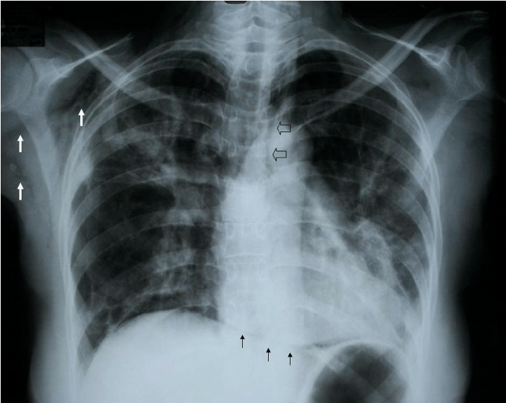
What Can Be Seen On A Normal Chest X
Normal chest X-ray shows normal size and shape of the chest wall and the main structures in the chest.
As described earlier, white shadows on the chest X-ray signify solid structures and fluids such as, bone of the rib cage, vertebrae, heart, aorta, and bones of the shoulders. The dark background on the chest X-rays represents air filled lungs. These lung fields are seen on either side of the heart and the vertebrae located in the center of the film.
Avoid Allergens And Illness
Exposure to allergens can cause the body to produce more mucus than normal. Allergens can include animal dander, pollen, dust mites, fragrance, certain foods, and much more. If you have allergies, avoiding these things can help reduce excess mucus production. Additionally, getting sick with a cold can cause mucus to build-up in your throat and sinus cavities. As the common cold season is approaching, wash your hands frequently, support your immune system, get enough sleep, and keep your distance from others.2
Also Check: What Helps Breathing With Asthma
You May Like: What Can Cause Asthma Exacerbation
Whats The Difference Between X
A chest X-ray is one method of providing your doctor with images of your heart and lungs. A computed tomography scan of the chest is another tool that is commonly ordered in people with breathing problems.
Unlike a standard X-ray, which provides a flat, one-dimensional picture, CT scans provide a series of X-ray images taken from different angles. It gives doctors a cross-section look at the organs and other soft tissue.
A CT scan gives a more detailed view than a regular X-ray. It can be used to check for blood clots in the lungs, which a chest X-ray cant do. A CT scan can also pick up much smaller detail, identifying problems, like cancer, much earlier.
The imaging test is often used to follow up any abnormalities seen within in the lungs on a chest X-ray.
Its not uncommon for your doctor to recommend both a chest X-ray and a CT scan depending on your symptoms. A chest X-ray is often done first because it is fast and accessible and provides useful information in order to make decisions quickly about your care.
How Many Chest X
We were unable to process your request. Please try again later. If you continue to have this issue please contact .
Every test has its risks as well as its benefits. I would only order a test if the results would change my clinical decision or management, and the same is true for x-ray studies. It is usually more common to order imaging studies during the initial evaluation of a patient with chronic respiratory symptoms, but once a diagnosis is established and treatment initiated, it is best to order studies much more selectively and based upon clinical changes or questions that come up. Personally, I think one chest x-ray is enough unless clinically indicated.
Figure 46-1. This is a chest radiograph from a 14-month-old girl with a 3-week history of cough, revealing a triangular density in the right hilar region and subtle air trapping in the right lower lobe. Rigid bronchoscopy with retrieval of a piece of glass from the right mainstem bronchus was performed and resulted in resolution of her symptoms.
Figure 46-2. Contrast-enhanced chest computed tomography image revealing a double aortic arch in a 3-year-old boy undergoing evaluation for chronic cough and wheeze, which had been persistent since early infancy. As part of his initial evaluation, a chest radiograph and barium swallow were performed, and these revealed a right-sided aortic arch. An MRI can also be performed to define vascular anatomy.
Suggested Readings
Don’t Miss: Can You Join The Air Force With Asthma
What Is A Ct Scan
CT stands for computed tomography. It uses X-rays to build a 3-dimensional picture of the inside of your body. This gives a detailed picture of your lungs, blood vessels and other organs.
You may be given an injection of material that shows up on the scan and can help to outline blood vessels. This is called contrast.
Does Asthma Show Up On Ct Scan
Ask U.S. doctors your own question and get educational, text answers â it’s anonymous and free!
Ask U.S. doctors your own question and get educational, text answers â it’s anonymous and free!
HealthTap doctors are based in the U.S., board certified, and available by text or video.
Don’t Miss: How To Get Rid Of Asthma Without Inhaler
Study Design And Setting
This was a retrospective cohort study conducted in Rochester, Minnesota, located in Olmsted County. The study protocol was approved by the Institutional Review Boards at both the Mayo Clinic and the Olmsted Medical Center. Medical care in Rochester is virtually self-contained within these two medical centers because, geographically, there are no other local medical clinic options. All inpatient and outpatient data have been indexed in an automated form since 1935 . Characteristics of the city of Rochester and Olmsted County populations were similar to those of the US Caucasian population, with the exception of a higher proportion of the working population employed in the healthcare industry.
What Are The Types Of Vocal Cord Dysfunction
- Laryngospasm: A laryngospasm is where your vocal cords seize or contract. You may lose your ability to speak and struggle to breathe. This can be caused by a reflux , environmental irritants and more.
- Exercised induced VCD: This is when the vocal cords move toward the middle when you are breathing during high intensity exercise, making it difficult to take air in.
- Irritant-induced VCD: Irritant-induced VCD is when the vocal cords contract with certain environmental triggers, such as strong scents, fumes, pollutants, chemicals and more.
- Stress-induced VCD: Vocal folds contract as a response to stress and anxiety.
Read Also: What To Do When Having An Asthma Attack
Recommended Reading: How To Make Asthma Go Away Without Inhaler
Identifying An Asthma Cough
The purpose of a cough is to remove foreign particles and bacteria to prevent a possible infection. There are two types of coughs: productive and nonproductive. When a cough is productive, it means that a noticeable amount of phlegm expelled. This enables the lungs to get rid of harmful substances.
Coughing in people with asthma can be helpful because its one of the bodys natural defense mechanisms. A productive asthmatic cough will expel phlegm and mucus from the lungs. In most cases of asthma, the cough is considered nonproductive. A nonproductive cough is a dry cough. Its a response to an irritant that forces the bronchial tubes to spasm . Swelling and constriction of the airways, which prompts this type of nonproductive cough, characterize asthma.
An asthma cough is also often accompanied by wheezing. This is a high-pitched whistling sound caused by a constricted airway.
What Are The Risks Of A Chest X
Chest X-rays expose the patient briefly to a minimum amount of radiation. Any radiation exposure has some risk to the tissues of the body. The radiation exposure in a chest X-ray is minimized by the type of X-ray high-speed film, which does not require as much radiation exposure as in the past. The radiology technician is guided by technique standards which have been established by national and international guidelines. These guidelines are designed and reviewed by both the Department of Health and Human Services and national and international radiology protection councils.
Women who are , especially in early , should notify their physicians, as the fetus is at risk for harm with any radiology technique. X-rays are typically avoided in pregnant patients unless absolutely necessary, in which case the patients abdomen is covered with a special lead gown to block the radiation from the fetus.
You May Like: J Asthma
Control Of Polymeric Mucin Production
The induction of Muc5ac in allergically inflamed mice is dependent upon two important signaling pathways: the IL-13/IL-4 receptor-α complex and the epidermal growth factor receptor . The functional dominance of these signaling pathways, however, does not translate into a simple intracellular pathway for Muc5ac gene activation. The principal signaling molecule activated by IL-13 is signal transducer and activator of transcription 6 . STAT6 signaling in mouse airway Clara cells is necessary and sufficient for Muc5ac induction and airway hyperreactivity in response to IL-13 . STAT6 binds to a canonical motif, 5â²-TTCN4GAA-3â², but this motif is not present in the conserved promoter regions of any mammalian MUC5AC orthologs . One indirect mechanism that may explain IL-13-mediated Muc5ac promoter activation is STAT6-dependent downregulation of forkhead box a2 . Foxa2 is a critical negative regulator of Muc5ac expression, and genetic deletion of Foxa2 in mice leads to constitutive Muc5ac overproduction resembling mucous metaplasia .
Transcriptional control of Muc5ac production gr2
Dont Miss: Is Eczema Related To Asthma
My Doctor Found A Spot On My Lung Now What
Getting answers and an explanation of whats causing the imaging results is of primary importance. You may be understandably concerned, and having your spot evaluated will give you the information you need.
At MXBowen, Physician, P.C., Health & Breathing Center, were here for you. Were here to provide diagnostic testing, specialist expertise, and peace of mind.
You May Like: Does Smoking Weed Help Asthma
What Happens During A Chest X
After you arrive at Northwest Pulmonary and Sleep Medicine, youll be ushered to a changing room. Youll typically need to undress from the waist up and remove your jewelry. Youll be provided with an exam gown to wear.
Depending on the images your practitioner has requested, you may have the X-ray while standing, sitting, or reclining. Heres what to expect next:
- Your technician moves you into position, usually a front or side position, or both
- Youll take a deep breath and hold it
- Youll need to stay very still, otherwise the image will be blurry
- The technician steps behind a window or wall during the X-ray
- You may be repositioned and repeat the process again
The technician makes sure youre comfortable throughout your X-rays, which are usually over quickly.
Afterward, your practitioner at Northwest Pulmonary and Sleep Medicine reviews the X-ray results with you. Together, youll discuss whether you need additional tests or treatment.
If youre experiencing breathing problems or have a chest injury and could benefit from a chest X-ray, call Northwest Pulmonary and Sleep Medicine today or book a consultation online.
- Chronic Obstructive Pulmonary Disease
- Common Cold
- Continuous Positive Airway Pressure
- Emphysema
- Idiopathic Hypersomnia – Sleeping 10+ hours
- Insect Sting Allergy
- Obesity & Sleep: Obesity Hypoventilation Syndrome
- Older Adults and Sleep Problems
- Pediatrics – Asthma
Symptom And Quality Of Life Scores
These tests involve filling out questionnaires to find out what symptoms youre experiencing and the different ways asthma may be affecting your life.
In an Asthma UK survey, 91% of people said their severe asthma had an impact on everyday things, such as their ability to exercise, their family and social life, their work or school life and their holidays. In the same survey 82% of people said theyd experienced sleep loss and 66% had gained weight.
If your asthma symptoms mean you cant do things you used to be able to do, such as exercise or play with your children, these scores will help your asthma specialist find out how asthma is affecting your life, says Dr Andy. Theyre also a useful way to monitor how well your asthma treatments are working.
Asthma specialists use these tests to:
- give them a clearer picture of your asthma symptoms
- show them how asthma may be limiting your activities
- reveal how youre coping with the challenges of having frequent asthma symptoms
- work out your response to longer-term treatment trials.
You May Like: What Happens If You Smoke Weed With Asthma
What If Its Not Copd
Chest discomfort can be caused by other conditions aside from COPD. If your chest X-ray doesnt show noticeable signs of COPD, your doctor will examine it for other possible issues.
Chest pain, difficulty breathing, and decreased ability to exercise can be symptoms of a lung problem, but they can also be signs of a heart problem.
A chest X-ray can provide valuable information about your heart and blood vessels, like heart size, blood vessel size, signs of fluid around the heart, and calcifications or hardening of valves and blood vessels.
It can also reveal broken ribs or other problems with the bones in and around the chest, all of which can cause chest pain.